
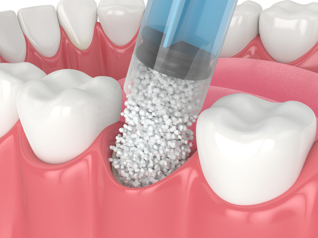
Dental bone grafting after a tooth extraction, clinically known as socket preservation, is a specialized procedure where biocompatible material is placed into an empty tooth socket. This material acts as a scaffold, stimulating your body to generate new jawbone, preventing your face from sagging. Clinical literature suggests that bone grafts are transplanted into a bony defect to promote healing and new bone formation [1] (Rajmalani et al., 2025).
What Are Dental Bone Grafts Made Of?
A common fear among patients is the belief that a bone graft requires a second, painful surgery to harvest bone from another part of the body. Today, socket preservation is a minimally invasive procedure that uses safe, lab-processed materials, completely eliminating the need for secondary surgeries. At Azalea Dental, we use highly regulated, sterile grafting materials that are 100% biocompatible with your body. Depending on your specific needs, Dr. Barbaro may use an allograft, a xenograft, or a synthetic graft. Medical literature supports that the decision to use a bone graft depends on the characteristics of the bone defect, the patient’s health, and the expected treatment outcome [2] (Zhang et al., 2025). Regardless of the type, these materials are stripped of all organic cellular matter. What remains is simply a sterile matrix, a microscopic framework that gives your own bone cells a physical structure to climb and rebuild upon.
Table that compares grafting options
Treatment approach | Cost impact | Durability | Future level of maintenance |
Extraction without a graft | Lowest upfront cost | Severe bone loss | Very high |
Extraction same-day | Highly affordable | Maximum bone preservation | Very low |
Delayed bone graft | Very high | Variable | High |
The Biological Timeline: How Your Body Builds New Bone
While your gum tissue will heal beautifully and close over the extraction site in just a few weeks, the bone regeneration happening underneath proceeds on a much slower biological timeline. Understanding this invisible process helps remove the anxiety of the waiting period before your permanent dental implant can be placed.
- Months 1 to 2: Immediately after the graft is placed, your body sends tiny blood vessels into the granular matrix. Clinical literature suggests that after tooth extraction, the socket fills with blood, forming a blood clot [3] (Udeabor et al., 2023).
- Months 3 to 4: Your cells begin absorbing the graft material, replacing it completely with your own natural bone tissue.
- Month 5 and Beyond: The temporary graft material has completely vanished, leaving behind a permanent foundation of your own jawbone, perfectly prepped for a titanium implant.
Upper vs. Lower Extractions: The Threat of Sinus Expansion
While losing bone density is a risk anywhere in your mouth, extracting an upper back molar can lead to sinus expansion. The roots of your upper back teeth actually push up into your maxillary sinus cavity, essentially acting like tent poles holding the sinus floor in place. When one of these upper molars is extracted and the “tent pole” is removed, the air-filled sinus cavity naturally begins to drop and expand downward into the empty space. This process is called sinus pneumatization. If this happens, you will lose the vertical bone height required to support a future dental implant, forcing you to undergo a much more invasive and expensive surgery later called a “Sinus Lift.” By placing a bone graft into that upper socket on the exact same day the tooth is pulled, we physically block the sinus from dropping, preserving your natural anatomy.
Biocompatibility: Is Socket Preservation Safe?
For our wellness-focused patients who closely monitor what goes into their bodies, the idea of foreign grafting material can prompt questions about safety and immune rejection. Because modern grafting materials are acellular, your immune system does not recognize them as a threat. In fact, systematic reviews suggest that each material has some efficacy in preserving the socket [4] (Alenazi et al., 2022). There is no risk of immune rejection like you might see in a major organ transplant. The material is thoroughly sterilized and regulated in accordance with strict FDA guidelines. Ultimately, the graft serves purely as a catalyst for your body’s natural healing mechanisms. It simply holds the space open, preventing your gums from caving into the hole, until your body’s natural metabolism replaces the minerals with your own living tissue.
3D Imaging: Removing the Guesswork from Bone Density
In the past, dentists had to rely on flat, two-dimensional X-rays to determine whether a bone graft had healed enough to support an implant. At Azalea Dental, we have eliminated that guesswork entirely by utilizing advanced dental technology. Before we ever clear you for the next phase of your tooth replacement journey, we utilize Cone Beam Computed Tomography (CBCT). This state-of-the-art scanner captures a high-definition, 3D replica of your jawbone in seconds. Instead of guessing, Dr. Barbaro can view your newly grafted bone from every conceivable angle on a monitor. We can measure the exact millimeters of the bone’s width, depth, and density.
FAQS
1. Do I really need a bone graft if I am getting a dental bridge instead of an implant?
Yes. Even if you plan to get a dental bridge, a bone graft is highly recommended. If you skip the graft, the bone under the bridge will eventually shrink and cave in, creating an ugly, visible gap between the bottom of your bridge and your gums, where food can easily get trapped.
2. How long does a dental bone graft take to heal before I can get an implant?
While your gums will close over the extraction site in about two to three weeks, the bone regeneration process happening underneath takes time. Most patients require 3 to 4 months for the graft to fully convert into dense, solid jawbone that is strong enough to support a permanent dental implant.
3. Does the bone grafting procedure add more pain to my extraction recovery?
Not at all. The bone graft is placed inside the empty socket while you are still completely numb from the extraction. Because the graft material protects the exposed bone and nerve endings, many patients actually report less post-operative sensitivity when a socket preservation graft is used.
References
[1] Rajmalani, M., Dadani, F., Iturbe, A., Kaur, J., Alla, O. W., & Mariela, N. (2025). Advances and applications of bone grafts in dentistry: A short review. Bioinformation, 21(12), 4687–4691. https://doi.org/10.6026/973206300214687
[2] Zhang, J., Zhang, W., Yue, W., Qin, W., Zhao, Y., & Xu, G. (2025). Research Progress of Bone Grafting: A Comprehensive Review. International journal of nanomedicine, 20, 4729–4757. https://doi.org/10.2147/IJN.S510524
[3] Udeabor, S. E., Heselich, A., Al-Maawi, S., Alqahtani, A. F., Sader, R., & Ghanaati, S. (2023). Current Knowledge on the Healing of the Extraction Socket: A Narrative Review. Bioengineering (Basel, Switzerland), 10(10), 1145. https://doi.org/10.3390/bioengineering10101145
[4] Alenazi, A., Alotaibi, A. A., Aljaeidi, Y., & Alqhtani, N. R. (2022). The need for socket preservation: a systematic review. Journal of medicine and life, 15(3), 309–312. https://doi.org/10.25122/jml-2021-0308