
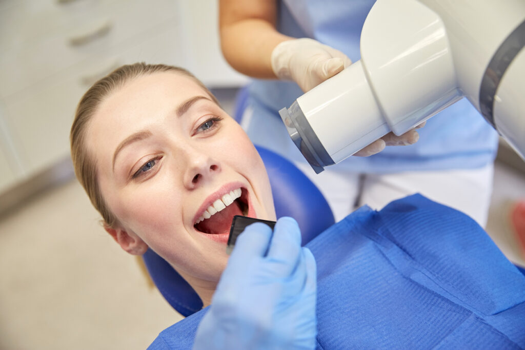
Digital dental X-rays are advanced diagnostic sensors used by dentists to capture high-definition images of teeth, jawbone, and surrounding soft tissues. Unlike traditional film, these digital systems at Azalea Dental use up to 90% less radiation and provide instant, high-contrast images that enable early detection of decay, bone loss, and infections.
Protecting Developing Smiles: Pediatric Digital X-Ray Standards
The medical literature supports the view that early detection of dental caries in children is critical to preventing irreversible tooth damage and ensuring optimal oral health outcomes [1] (Rahimi et al., 2026). Pediatric dental X-rays are a specialized preventive tool used to monitor the growth of adult teeth and ensure the jaw is developing correctly. Because children’s tissues are more sensitive to radiation, our use of digital sensors is vital. These sensors require a fraction of the exposure time used in traditional clinics. We typically use “Bitewing” X-rays for children to detect cavities early in baby teeth, which helps prevent premature tooth loss and orthodontic issues. By keeping exposure 90% lower than old-fashioned film, we ensure your child’s visit to our Wilmington office is both medically thorough and biologically safe.
A table that compares X-rays
X-ray type | Primary purpose | Frequency | Maintenance level |
Bitewings | Detecting hidden cavities | 6-12 months | Routine prevention |
Periapical | Root health and infections | As needed | Diagnostic/restorative |
Panoramic | Wisdom teeth and orthodontic planning | 3-5 years | Developmental |
CBCT | Implant placement and bone density | Pre-surgical | Advanced restorative |
Detecting Bone Loss and Hidden Infections
Nobody wants to invest their hard-earned money in a cosmetic smile makeover only for it to fail months later because of hidden decay. If you are hoping for a brighter, straighter smile for that upcoming promotion or wedding, your comprehensive exam is step one. Before we can safely recommend professional teeth whitening, porcelain veneers, or clear aligners, we must ensure your jawbone and enamel are structurally sound. Your baseline visit gives us the exact green light we need to safely design the flawless, camera-ready smile you deserve without any structural surprises down the road.
When You Need a CBCT Scan
Sometimes, a flat 2D image is not enough to provide a safe diagnosis. Clinical evidence suggests that CBCT imaging has overcome the limitations of 2D imaging, offering practitioners high-quality, sub-millimeter-resolution images with short scanning times and low radiation doses [3] (Venkatesh et al., 2017). For patients preparing for dental implants or wisdom tooth removal, we utilize Cone Beam Computed Tomography (CBCT). A CBCT scan is a specialized digital X-ray that creates a 3D model of your entire head and jaw. This allows us to see exactly where your facial nerves and sinus cavities are located. By virtually “practicing” your surgery on a 3D digital model before we ever pick up a tool, we minimize your time in the chair and maximize your surgical safety. This level of technology is ideal for restorative success and is a favorite for our patients who value precision and efficiency.
Faster Insurance Approvals and Specialist Coordination
One of the “hidden” benefits of digital X-rays is the speed of administrative processing. Because digital files are captured instantly, our team can electronically attach your high-definition images to insurance claims for Delta Dental or Blue Cross Blue Shield. This often leads to faster approvals and fewer “denied” claims for major work, such as crowns or bridges. Furthermore, if you need a referral to a local Wilmington specialist, we can securely email your X-rays within seconds, eliminating the need to pick up physical film or pay for duplicate scans at another office.
Seeing Your Smile in 4K
With digital X-rays, we practice “Co-Diagnosis.” We project your high-contrast images onto a large 4K monitor right in front of your chair. This allows you to see exactly what Dr. Barbaro sees. We can zoom in on a microscopic fracture or use color-shading tools to show you exactly where a cavity is starting. When you can see the problem for yourself, the “mystery” of dental work disappears. You become an active partner in your healthcare decisions, ensuring your treatment plan perfectly aligns with your goals.
FAQS
1. Are digital dental X-rays safe during pregnancy?
Yes. While we minimize elective imaging during pregnancy, the ADA and ACOG confirm that diagnostic X-rays with proper lead shielding (including a thyroid collar) are safe. We prioritize treating active infections to protect both mother and baby.
2. Why do I need X-rays if my teeth don't hurt?
Dental pain usually occurs only after decay has reached the nerve. Digital X-rays allow Dr. Barbaro to see “micro-cavities” and early bone loss while they are still treatable with simple, low-cost fillings rather than expensive root canals.
3. How long does the X-ray process take?
Digital sensors are nearly instantaneous. Unlike the 10-minute wait for film development, these images appear on our chairside monitors within seconds, allowing you to see exactly what the doctor sees in real time.
References
[1] Rahimi, H., Naeimi, S. M., Darvish, S., Nazemi Salman, B., Razzaghi, P., Luchian, I., & Budala, D. G. (2026). Detection of Pediatric Dental Caries in Panoramic Radiograph Using Deep Learning: A Benchmark Study on MD-OPG. Sensors, 26(8), 2481. https://doi.org/10.3390/s26082481
[2] Graham J. (2015). Detecting low bone mineral density from dental radiographs: a mini-review. Clinical cases in mineral and bone metabolism : the official journal of the Italian Society of Osteoporosis, Mineral Metabolism, and Skeletal Diseases, 12(2), 178–182. https://doi.org/10.11138/ccmbm/2015.12.2.178
[3] Venkatesh, E., & Elluru, S. V. (2017). Cone beam computed tomography: basics and applications in dentistry. Journal of Istanbul University Faculty of Dentistry, 51(3 Suppl 1), S102–S121. https://doi.org/10.17096/jiufd.00289